Like the film inside of a camera, the retina is responsible for processing the images projected onto it and then the optic nerve transmits this to the brain. The retina is susceptible to many types of diseases and the specially trained eye doctors at Grand Rapids Ophthalmology are here to help you! They will diagnose and treat a variety of retinal conditions including macular degeneration, diabetic retinopathy, ocular histoplasmosis, retinal detachment and more.

A thin layer of tissue called the retina covers the back inside wall of the eye. When the cornea and lens at the front of the eye focus light on the retina, a picture is taken and sent to the brain through the optic nerve to be interpreted. Thus, the retina is the “seeing tissue” of the eye.
The retina has two parts: the peripheral retina and the macula.
The macula is the center of the retina, while the large area surrounding the macula is the peripheral retina. The peripheral retina allows us to see out of the corner of the eye but is unable to see detail clearly. We cannot drive, read or see facial features with the peripheral retina.
Although small, the macula is one hundred times more sensitive to detail than the peripheral retina. The macula is required to see fine detail and it must be healthy to work properly.
The vitreous is a jelly-like substance that fills the cavity between the lens and the back of the eye. Also called the vitreous humor. As we age, the vitreous ages along with the rest of the body, and these aging changes can sometimes lead to problems with vision and even damage to the retina. That is why we often speak of patients with vitreo-retinal disease.
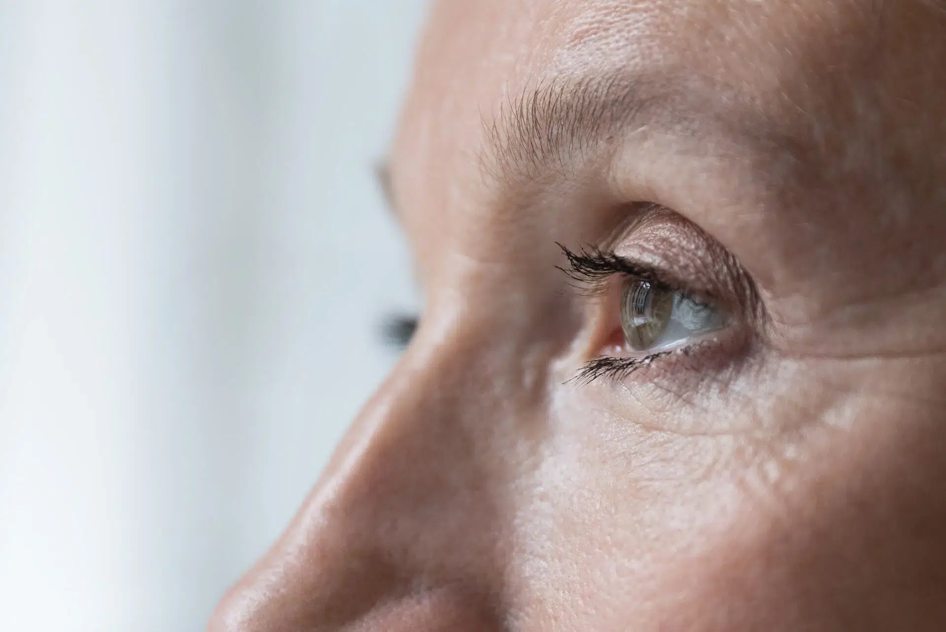
After you complete your paperwork with a receptionist, a technician will perform an initial assessment. Please bring your glasses and/or contact lenses with you so that your vision can be measured with them in place. The technician will obtain your history, discuss your specific concerns and apply numbing drops and check the pressure in your eyes. A second set of drops will be given to dilate (widen) your pupils. Dilation is necessary for a complete examination of the inside of your eyes. Your pupils may require up to 30 minutes to dilate.
Since your pupils may remain dilated for a full day after your visit we recommend that your bring sunglasses to reduce sensitivity to light. Your ability to read will be temporarily decreased. Although distance vision is usually undisturbed, we recommend a driver if possible.
After your eyes are dilated you will see the doctor for the eye examination. A final opinion may not be possible until all test results are analyzed. Your doctor will usually discuss the available findings with you on the day of your visit and initiate treatment if necessary. Following careful studies, reports will be forwarded to your personal physician(s).
Please bring a list of prescription medications you are currently taking, including the dosage. We thank you in advance for giving us the most complete information possible. Family members and friends are welcome and encouraged to accompany you to your appointment.
Age-related macular degeneration (AMD) is a common eye condition and a leading cause of vision loss among older adults. It is caused by the thinning of the macular tissue and can cause central vision loss, blind spots, changes in color perception, and distorted vision.
Diabetes is a chronic condition that affects the body’s ability to process blood sugar. People with diabetes are at risk for diabetic retinopathy, an eye disease that damages the smallest retinal blood vessels. Diabetic retinopathy has several distinct stages with varying symptoms. Early detection is important to prevent permanent vision loss. Learn more about diabetic retinopathy and available treatment options at Grand Rapids Ophthalmology on our Diabetic Eye Disease page.
Flashes and floaters are common visual disturbances that appear as flashes of light or as dark specks or strands. Flashes and floaters are typically the result of changes in the vitreous gel but can also be signs of more serious disease. They are often harmless, but it is important to have them evaluated by an experienced eye doctor if you experience this type of visual disturbance.
The macula is delicate, and if it becomes swollen or inflamed, a condition called macular edema can occur. This condition causes decreased central vision and may also cause redness and discomfort. Macular edema is often associated with other retinal diseases and can be treated with eye drops, injectable medications, and sometimes surgery.
A macular hole occurs when the vitreous pulls away from the retina in a way that causes a gap to form in the macula. A macular hole causes blurriness in central vision, makes straight lines look wavy, and creates difficulty reading and performing daily tasks. Surgery may be used to repair a macular hole.
Shrinking of the vitreous is a natural part of the aging process and can lead to the development of scar tissue that may cause the macula to wrinkle or pucker. This condition typically causes symptoms such as mild blurriness or distortion in vision. If a macular pucker does not resolve on its own and it causes significant symptoms, it can be treated with surgery.
A retinal tear can occur due to aging or an eye injury. Similar to retinal detachment, a tear in the retina can cause shadows in vision or flashes. If left untreated, a retinal tear can lead to retinal detachment. Retinal tears often require treatment but not always.
A detached retina occurs when the retina separates from the underlying layers of the eye, which can cause symptoms including vision loss, sudden shadows, bright flashes, and floaters. Retinal detachment can happen gradually or suddenly and can be associated with inflammatory conditions, vitreous aging changes, or an eye injury. Retinal detachment is a medical emergency and should be diagnosed immediately. A variety of surgical techniques can be employed to treat retinal detachment, and retinal specialists will discuss and offer the best alternatives suited to your specific details. The timing of retinal detachment repair can range from emergent (within a day or two) to within many weeks or even months.
The small blood vessels of the retina transfer nutrients and oxygen to and from the eye and are essential for proper retinal function.
If a retinal vein becomes blocked, it causes retinal vein occlusion (RVO). Patients with RVO may have sudden, painless loss of vision in one eye. The treatment for RVO depends on the type, location, and severity of the blockage in the retinal vein and may consist of observation, injections of medication, and sometimes surgery. It is very important for people with risk factors such as inflammatory eye conditions, high blood pressure, cardiovascular, glaucoma, or diabetes to receive regular eye examinations and preventive care.


